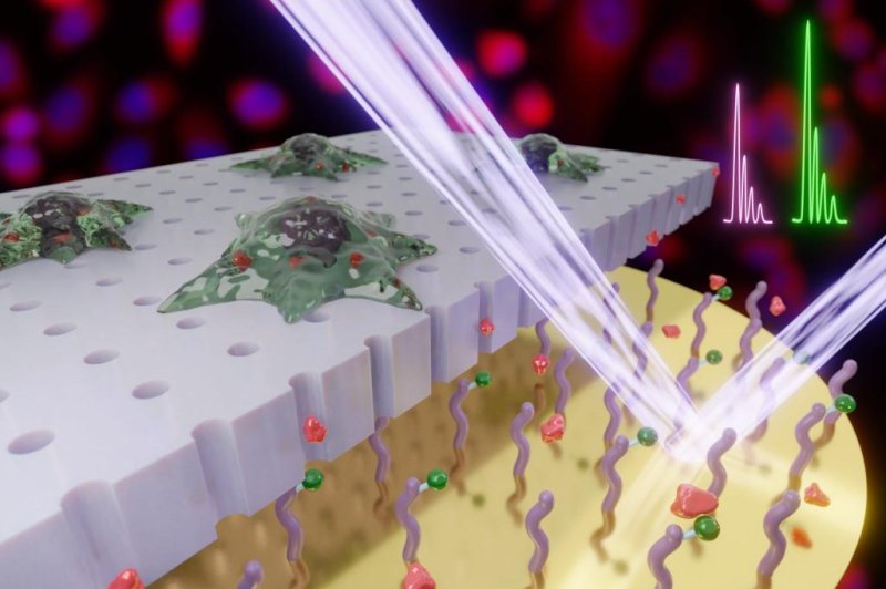
Engineers at Northwestern University have developed a method for extracting multiple samples from a single cell over time without causing permanent damage. Photo by Horacio D. Espinosa, Prithvijit Mukherjee, Eric Berns, and Milan Mrksich
May 26 (UPI) -- Scientists have developed a new method for sampling cells multiple times without causing permanent damage to the cell.
Most cell sampling and analysis methods, including genetic and protein sequencing, destroy the target cell. As a result, sampling results provide only a single snapshot.
Cells are complex and dynamic. Capturing their evolving behaviors and their reactions to outside stimuli requires more than a snapshot frozen in time.
The new method, called localized electroporation, uses mass spectrometry to sample and analyze enzymatic activity inside a cell without doing irreparable harm. The technique relies on what scientists dubbed the live cell analysis device, or LCAD. The device allows scientists to perform what is essentially a biopsy, but at nano scales on a single cell.
Localized electroporation and the LCAD, described Monday in the journal Small, could be used to study a variety of cellular behaviors and reactions. For example, the novel technique could be used to observe how cells respond to different cancer treatments.
"By exploiting advances in microfluidics and nanotechnology, localized electroporation can be employed to temporarily open small pores in the cell membrane enabling the transport of molecules into the cells or extraction of intracellular contents," study co-author Horacio Espinosa, professor of manufacturing and entrepreneurship at Northwestern University's McCormick School of Engineering, said in a news release. "Since the method is minimally invasive to the cells, it can be repeated multiple times without their disruption."
"Certain enzymes may be linked to disease pathways, such as certain types of cancers, and they may be the target of therapeutics," said study co-author Milan Mrksich, Northwestern University vice president for research. "Using this platform, it is now possible to study how enzymatic activity varies between healthy cells and cells from a tumor biopsy."
The technique could also be used to study how a cell's enzymatic activity responds to different types of treatment.
Instead of lots of single snapshots, scientists will getting the equivalence of a motion picture of cellular changes, allowing researchers to study a variety of dynamic cellular processes, including cell differentiation, disease progression and drug response.
"We envision that this technique can be used in scenarios such as screening drugs or designing and optimizing treatment courses that can arrest disease progression in cells," Espinosa said.
In addition to delicately and precisely extracting cellular material, the LCAD could be used to deliver new materials, like edited DNA or proteins, to a cell.
"We have used the same concept of localized electroporation to do CRISPR gene editing and we are now using machine learning to automate the process," Espinosa said.


