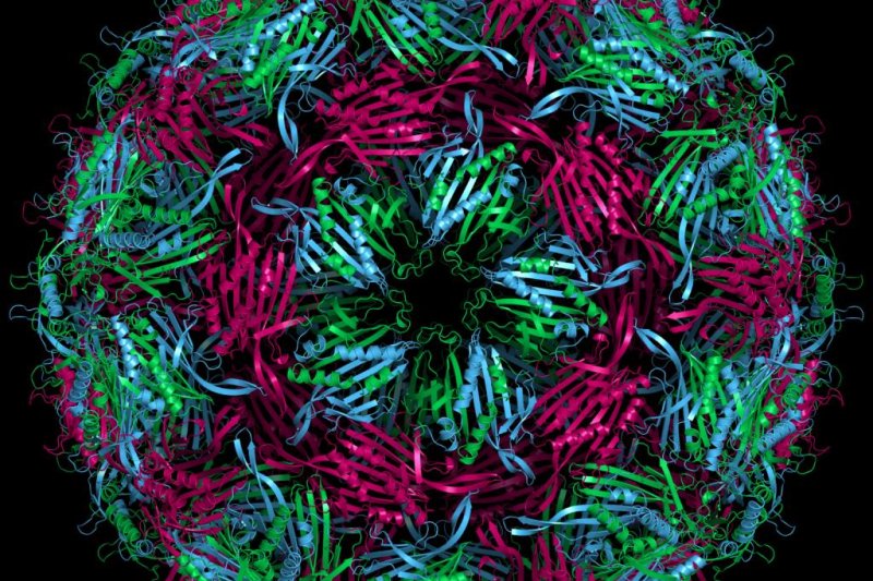
Scientists used a novel imaging technology to study the formation of bacteriophage MS2, a virus that infect E. coli bacteria. Photo by
Wikimedia Commons
Oct. 4 (UPI) -- Scientists have captured images of individual viruses forming, gaining insights into the mechanics of viral assembly.
"Structural biology has been able to resolve the structure of viruses with amazing resolution, down to every atom in every protein," Vinothan Manoharan, a professor of physics and chemical engineering at the Harvard University, said in a news release. "But we still didn't know how that structure assembles itself. Our technique gives the first window into how viruses assemble and reveals the kinetics and pathways in quantitative detail."
For the study, Manoharan and his research partners focused on the bacteriophage MS2, a virus that infects E. coli bacteria. MS2 is a single-stranded RNA viruses, the most abundant type of virus on Earth, and the type responsible for a variety of human maladies, including West Nile fever, gastroenteritis, hand, foot and mouth disease, polio, and the common cold.
Like most RNA viruses, the bacteriophage MS2 is relatively simple. It consists of a single piece of RNA with a diameter of 30 nanometers, boasting 3,600 nucleotides and 180 identical proteins. The proteins form hexagons and pentagons and organize themselves into a soccer ball-like shell, or capsid, around the virus.
To watch this formation process in real-time, scientists used interferometric scattering microscopy, a light scattering-based imaging technique. When light is scattered off the target object, it creates a dark spot in a larger field of light, which scientists observe as a proxy for the target itself.
In the lab, scientists watched as light scattered off RNA strands on a substrate created larger and larger dark spots in the larger field of light. Researchers were able to determine how many proteins were attaching to each RNA strand by watching the changing intensity of the dark spots.
"One thing we noticed immediately is that the intensity of all the spots started low and then shot up to the intensity of a full virus," Manoharan said. "That shooting up happened at different times. Some capsids assembled in under a minute, some took two or three, and some took more than five. But once they started assembling, they didn't backtrack. They grew and grew and then they were done."
Using models, scientists have previously hypothesized that viruses form one of two ways. One possibility is that proteins attach themselves sporadically to RNA strands and then organize to form a capsid. A second scenario requires a critical mass of proteins, or nucleus, to aggregate before the capsid can be formed.
The latest research -- detailed this week in the journal PNAS -- showed the second scenario best explains how a virus forms.
The timing of nucleus formation varies among different viruses, but the latest findings suggest that once a critical mass of proteins is reached, the virus begins to grow more quickly until it is fully formed.
Experiments in the lab showed that when a substrate was overrun with proteins, the virus was more likely to be improperly assembled.
"Viruses that assemble in this way have to balance the formation of nuclei with the growth of the capsid," said Manoharan. "If nuclei form too quickly, complete capsids can't grow. That observation might give us some insights into how to derail the assembly of pathogenic viruses."
The world's first video of assembling viruses fails to explain exactly how proteins organize themselves into capsids, but it does confirm the basic pathway for viral formation.


