Scientists say the new imaging technology could be used to study a variety of nanoparticles. Photo by Martin Gruebele/AIP
Feb. 8 (UPI) -- For the first time, scientists have managed to image electronically excited quantum dots in 3D. Quantum dots are tiny semiconductor particles, measuring just a few nanometers across.
The breakthrough promises to improve scientists' understanding of the unique nanoscale particles and their potential for a variety of technological applications.
In imaging quantum dots, scientists were keen to understand how potential defects in the semiconductor materials that yield the nanoparticles affect their quantum properties and behavior.
"Understanding how the presence of defects localizes excited electronic states of quantum dots will help to advance the engineering of these nanoparticles," Martin Gruebele, a physicist at the University of Illinois at Urbana-Champaign, said in a news release.
Defects are mostly avoided in material science, but they are welcomed in quantum technologies. Defects can help scientists tailor quantum dots for specific applications.
"Missing atoms in a quantum dot or substituting a different kind of atom are defects that will alter the electronic structure and change the semiconductivity, catalysis or other nanoparticle properties," Gruebele said. "If we can learn to characterize them better and precisely control how they are produced, defects will become desirable dopants instead of a nuisance."
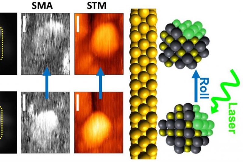
To image the quantum dots, researchers used an imaging technique called single molecule absorption scanning tunneling microscopy, or SMA-STM. The technology was first developed by Gruebele in 2005.
Scientists used a laser to roll a single quantum dot onto the wire tip of the microscope, allowing the imaging technology to capture slices of the dot at different orientations. The slices are then combined to build a 3D image of the nanoparticle.
Researchers are now conducting follow-up tests to ensure the quantum dot isn't damaged is it is rolled into each different orientation.
"We speculate that, in the future, it may be possible to do single-particle tomography if damage to quantum dots can be avoided during repeated manipulation," Gruebele said.
The research was published this week in the Journal of Chemical Physics.


