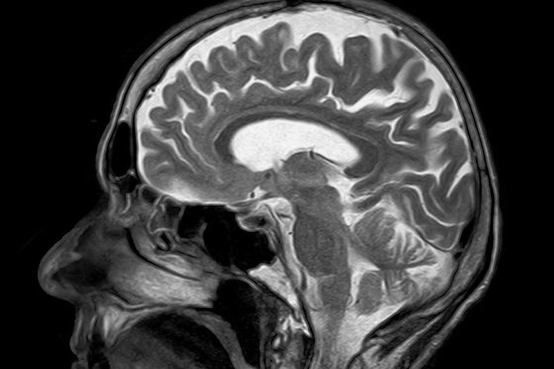
More than 22,000 Americans are diagnosed with glioblastoma annually. Photo by
toubibe/Pixabay
Feb. 6 (UPI) -- A new, more aggressive surgical approach may double survival rates in adults with brain tumors, a study published Thursday in the Journal JAMA Oncology has revealed.
The new procedure involves the removal of the tissue surrounding the tumor in glioblastoma, the most common -- and deadly -- type of brain cancer. This tissue is called the "non-contrast-enhancing tumor" because it does not appear on MRI exams even when a contrast agent is injected into the vein.
"Traditionally, the goal of neurosurgeons has been to achieve total resection, the complete removal of contrast-enhancing tumor," co-author and neurosurgeon Mitchel Berger, director of the University of California-San Francisco Brain Tumor Center, said in a press release. "This study shows that we have to re-calibrate the way we have been doing things and, when safe, include non-contrast-enhancing tumor to achieve maximal resection."
More than 22,000 Americans are diagnosed with glioblastoma annually. The cancer is perhaps best known for claiming the lives of senators John McCain and Edward Kennedy, as well as Beau Biden, son of former vice president and current presidential candidate Joe Biden.
Historically, survival rates have generally been lower for those with glioblastomas featuring so-called "IDH-wild-type" genetic mutations when compared to those with "IDH mutant" tumors.
For their research, Berger and his colleagues tracked 761 newly diagnosed glioblastoma patients at UCSF who were treated between 1997 and 2017, 62 of whom had IDH-mutant tumors or were under 65 years of age with IDH-wild-type tumors and were treated with both radiation and chemotherapy using temozolomide, a medication commonly used against glioblastoma.
Each also underwent surgeries in which a median of 100 percent of contrast-enhancing tumor and a median of 90 percent of non-contrast-enhancing tumor were removed. Notably, these patients survived an average of 37 months, or 3.1 years, following diagnosis.
Conversely, their counterparts -- 212 patients under 65 years of age who had received the same therapies but had more modest resections of the non-enhancing tumor -- survived only 16.5 months, or 1.4 years, on average -- about half as long.
Among the group of longer-surviving patients, those with IDH-wild-type tumor did approximately as well as those with the IDH-mutant variant when a portion of the non-contrast enhancing tumor was removed, the authors noted.
"The difference was that the patients with IDH-wild-type tumor declined more rapidly after the three-year mark," said co-author Annette Molinaro, from the UCSF Department of Neurological Surgery.
As promising as these findings are, the researchers caution that removal of the non-contrast-enhancing tumor should only be performed with techniques such as intra-operative brain mapping. This means that areas of the brain responsible for speech, motor, sensory and cognition are tested during surgery to ensure that they can be preserved.
The authors noted that approximately 80 percent of medical centers do not currently offer brain mapping.


