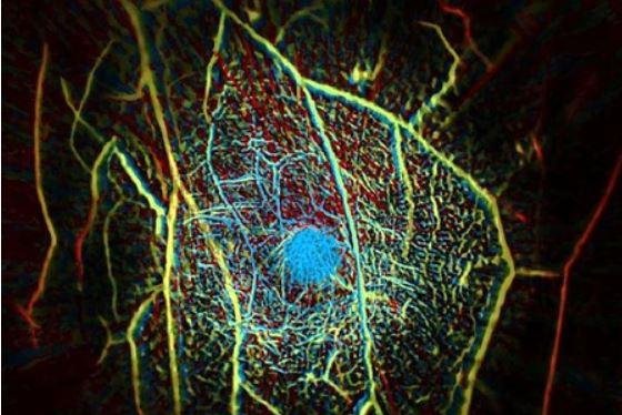
This is an image of the internal vascular structure of a human breast created with a new scanner designed at Caltech. Photo courtesy of California Institute of Technology
June 18 (UPI) -- Researchers said they developed a quicker, better and cheaper way than mammograms to detect breast cancer: A laser scanner that can find tumors in as little as 15 seconds.
The near-infrared lasers shine pulses of light into the breast tissue, which, researchers report in a study published Friday in the journal Nature Communications, is more effective than traditional mammograms.
"Our goal is to build a dream machine for breast screening, diagnosis, monitoring and prognosis without any harm to the patient," Lihong Wang, a professor of Caltech's medical engineering and electrical engineering, said in a press release. "We want it to be fast, painless, safe and inexpensive."
Wang is a founder of a company licensed to develop the system for large-scale clinical studies and commercialization of the technique.
With about 1 in 8 women in the U.S. developing invasive breast cancer during their lifetime, Wang said mammograms are valuable for reducing breast cancer deaths. But he said women are exposed to X-ray radiation and the method requires their breasts to be painfully flattened between plates for a clearer image.
Wang cited an analysis of 10 databases in 2013 where 25 percent to 46 percent of the women avoided mammograms because of pain as another strong reason that other ways to screen for breast cancer are needed.
Additionally, mammography doesn't accurately detect "radiographically dense" breasts -- usually in young women -- because they are opaque to X-rays. And mammography tests in half the cases generate false-positive diagnosis at some point in their lives.
MRI scans, which are expensive, sometimes require agents to be injected into the patient's blood, which can cause nausea and vomiting.
The new system is known as photoacoustic computed tomography, or PACT.
The patient lies face down on a table with a recess containing ultrasonic sensors and the laser. One breast at a time is placed in the recess.
"This is the only single-breath-hold technology that gives us high-contrast, high-resolution, 3-D images of the entire breast," Wang said.
This process takes about 15 seconds, compared to 45 minutes for MRI scans.
By shining a near-infrared laser pulse into the breast tissue, the laser light diffuses through the breast and is absorbed by oxygen-carrying hemoglobin molecules in the patient's red blood cells. And by causing the molecules to vibrate ultrasonically, they travel through the tissue and are picked up by 512 tiny ultrasonic sensors around the skin of the breast.
The process assembles an image of the breast's internal structures similar to ultrasound imaging, but is more precise -- as small as a quarter of a millimeter at a depth of 4 centimeters.
The images also show blood vessels present in the tissue being scanned, which can find tumors that grow because of the large amounts of blood.
Researchers tested the system on eight women and correctly identified eight of the nine breast tumors present.
In addition, Wang said PACT can be used on other parts of the body, including assessing the health of the vascular tissue in the extremities of a patient with diabetes.


