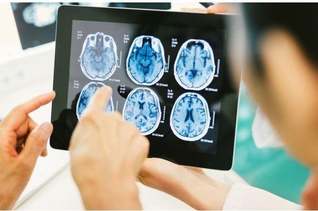
Medical personnel look at brain scans of patients with traumatic brain injuries. Photo courtesy of Walter Reed National Military Medical Center
April 30 (UPI) -- By finding changes to tiny blood vessels in the brains of people with traumatic injuries, researchers believe medical personnel could be able to make more precise diagnosis and treatment decisions.
Researchers found that changes in the blood vessels may be linked to a range of cognitive symptoms after a traumatic brain injury, or TBI. The research, which was led by the University of Pennsylvania, was presented Friday at the American Academy of Neurology Annual Meeting and has not yet been published.
"The relationship between microvascular and structural injury in chronic TBI has been recognized for years, but underappreciated," Dr. Ramon Diaz-Arrastia, director of the Traumatic Brain Injury Clinical Research Center at the Perelman School of Medicine at Pennsylvania, said in a press release. "This research adds another layer to our understanding of TBI and ways to better treat patients, who in some cases have had TBI symptoms for years."
Traumatic brain injury annually accounts for 2.5 million emergency department visits, 280,000 hospitalizations and nearly 50,000 deaths, according to the Centers for Disease Control and Prevention. TBIs contribute to about 30 percent of all injury deaths
The researchers wanted to assess the correlation between blood flow and cerebral reactivity, which is a change in blood flow in the brain in response to an injury.
They assessed the strength and function of small blood vessels in the brains of 27 chronic TBI patients and 14 healthy people. The researchers were able to see inside their brains by using the functional MRI-Blood-Oxygen Level dependent, or BOLD, and Diffusion Tensor Imaging, or DTI.
In addition, seven neuropsychological tests were performed with the Brief Symptom Inventory-Somatic and Rivermead Post-Concussion Questionnaire. It noted the severity of cognitive and emotional symptoms, including headaches and depression.
The researchers found differences in several regions of the brain, including deficits in cerebrovascular reactivity with TBI patients. Although vascular reactivity is decreased in chronic TBI, they found increased cerebrovascular reactivity in subcortical regions of the brain -- hippocampus, amygdala, thalamus, caudate, putamen. They are associated with post-concussive symptoms.
Diaz-Arrastia said the research provides hope for better treatment of TBI, and possibly other disorders, should assessment of neurological microvascular function becomes part of routine examinations -- which will help determine courses of treatment.
"These findings underscore the importance for precise diagnosis with TBIs, to ensure the right therapies are identified for patients," Diaz-Arrastia said. "By nature, TBI injuries always vary -- the brain damage is not the same in any two patients. If we have a therapy that could target the specific lesion that's unique for each patient, then we can treat patients with better, more appropriate therapies."


