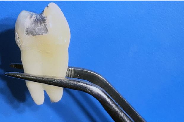
Researchers have found amalgam fillings exposed to ultra-high-strength MRI scans may release toxic mercury, according to a study. Photo courtesy of Radiological Society of North America
June 27 (UPI) -- When amalgam fillings, sometimes referred to as silver fillings, are exposed to ultra-high-strength MRI scans, they can release toxic mercury, according to a new study.
While researchers found no effects from the 1.5 Tesla MRI scans, which are lower strength and used commonly, the 7.0 Tesla, or T, version approved by the U.S. Food & Drug Administration in 2017 may have an unforeseen risk for people with amalgam fillings. The findings were published Tuesday in the journal Radiology.
The amalgam filling, which includes 50 percent mercury, can cause health problems. The FDA has said amalgam fillings are safe for adults and children older than 6, compared to in Sweeden, Norway, Denmark and Germany, where they have been restricted or forbidden.
"In our study, we found very high values of mercury after ultra-high-field MRI," Dr. Selmi Yilmaz, a dentist and faculty member at Akdeniz University in Antalya, Turkey, said in a press release. "This is possibly caused by phase change in amalgam material or by formation of microcircuits, which leads to electrochemical corrosion, induced by the magnetic field."
Previous research has found that exposure to the magnetic fields associated with MRI scans can cause mercury to leak from amalgam fillings, but the newer 7-T scanners had not been studied for their potential effects, researchers say.
For the study, Yilmaz and her colleague, Dr. Mehmet Zahit Adien, studied mercury released from dental amalgam after 7-T and 1.5-T MRI in teeth that had been extracted from patients for clinical indications.
They opened two-sided cavities in each tooth and applied amalgam fillings.
Nine days later, two groups of 20 randomly selected teeth were placed in a solution of artificial saliva followed by 20 minutes of exposure to 1.5-T or 7-T MRI. Artificial saliva was used in a control group.
The mercury content in the 7-T group, at .67 parts per million, was approximately four times the levels found in the 1.5-T group and the control group.
"In a completely hardened amalgam, approximately 48 hours after placing on teeth, mercury becomes attached to the chemical structure, and the surface of the filling is covered with an oxide film layer," Yilmaz said. "Therefore, any mercury leakage is minimal."
While Yilmaz said it is unclear how much of this mercury is actually absorbed by the body, adding that patients with amalgam fillings don't need to be all that concerned about have a 1.5-T MRI.
She said the researchers are undergoing three projects to examine phase and temperature changes of dental amalgam in different magnetic fields, in addition to looking into additional studies on MRI and the release of mercury from dental amalgam.


