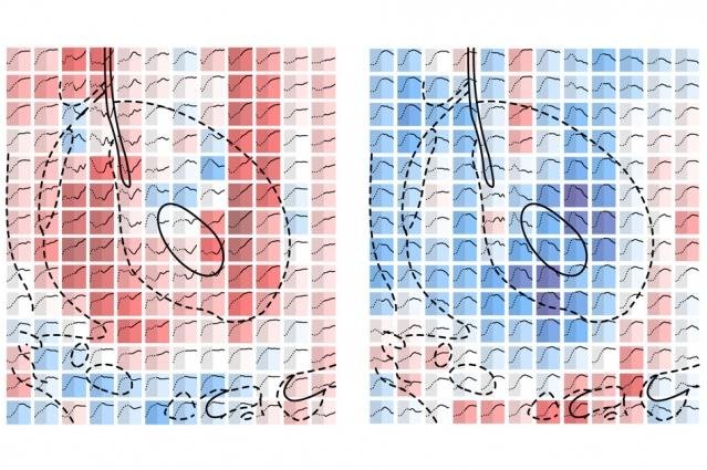
MIT researchers have imaged serotonin dynamics in a living brain. Pictured, molecular fMRI data showing signal changes from serotonin sensors in the absence (left) and presence (right) of the antidepressant Prozac Red squares indicate the signal has increased, as more serotonin is absorbed into neurons; blue squares indicate the signal has decreased, as less serotonin is absorbed into neurons. Dotted and solid lines graphed in each square show how the signal changes over time. The swirling black lines indicate features of the brain. A computer model uses this data to estimate neurotransmitter reuptake across the brain. Photo courtesy MIT
CAMBRIDGE, Mass., Oct. 20 (UPI) -- Researchers at the Massachusetts Institute of Technology have developed a three-dimensional imaging technique that maps serotonin as it's reabsorbed into neurons in a living brain.
Serotonin is a neurotransmitter associated with positive feelings such as happiness in humans. Due to its euphoric properties, the neurotransmitter is often targeted by antidepressants, which block serotonin receptors. MIT researchers say their first-of-its-kind technique may help make drugs more effective.
"Until now, it was not possible to look at how neurotransmitters are transported into cells across large regions of the brain," lead researcher Aviad Hai in a press release. "It's the first time you can see the inhibitors of serotonin reuptake, like antidepressants, working in different parts of the brain, and you can use this information to analyze all sorts of antidepressant drugs, discover new ones, and see how those drugs affect the serotonin system across the brain."
When researching the effects of antidepressants, scientists typically use an approach called microdialysis, which involved inserting a probe into the brain to collect tiny chemical samples from the tissue. In their 3D mapping project, MIT researchers sought to streamline this process by making it quicker and more accurate.
In the new technique outlined in a paper published in the journal Neuron, the research team engineered a protein that directly attaches itself to serotonin, and detaches during reuptake. The protein then produces a signal that can be analyzed using functional magnetic resonance imaging, or fMRI.
"Basically, what we've seen in this work is a method for measuring how much of a neurotransmitter is being [absorbed], and how that amount, or rate, is affected by different drugs ... in a highly parallel fashion across much of the brain," author Alan Jasanoff explained.
While testing their technique, scientists recorded a stronger decrease of serotonin reuptake with the antidepressant drug Prozac. Authors of the study say their findings provide more proof of the strong relationship between serotonin and dopamine systems.


