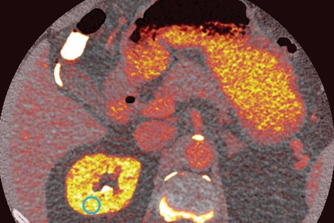
Greater amounts of iodine contrast are shown in brighter, yellow colors in a scan of a research subject at the NIH Clinical Center taken during tests of a new photon-counting CT scanner. Photon CT scan images may offer doctors better views for diagnosis and treatment. Photo by National Institutes of Health
BETHESDA, Md., Feb. 24 (UPI) -- A new type of computerized tomography, or CT, scanner being tested by the National Institutes of Health could give doctors much more detailed views inside the body while exposing patients to far less radiation.
The photon-counting detector CT scanner is currently just a prototype, but the new imaging device is expected to provide detail down to half a millimeter and allow for better identifications of tissues.
The NIH is working with Siemens Healthcare to test the new device at the agency's clinical center in Bethesda, Md., where agency researchers said the device can be tested in the diagnosis and treatment of rare and complicated illnesses as they design protocols and methods for its use.
"The NIH Clinical Center has helped shape and share research advances and health care for decades," Dr. David Bluemke, chief of the Department of Radiology and Imaging Sciences at the Clinical Center, said in a press release. "Now is an exciting time for us and for our study participants here in the Clinical Center as we help test and develop this CT technology so that it may one day help patients around the world and impact the health care they receive."
Researchers recruited 15 asymptomatic volunteers with a mean age of 58.2 years, seven of whom were men, between September and November of 2015 to undergo scans in the machine.
Scans showed no statistically significant difference in image quality or usefulness over traditional scans, though the new scanner offered additional spectral information researchers said may prove to be useful.
The first findings from tests of the scanner were published in the journal Radiology.
Future tests, researchers said, will focus on three areas: more detailed pictures using contrast dyes for faster and more accurate diagnosis; attempting to identify tumors, plaques, and vessels smaller than half a millimeter; and more accurately identifying soft tissues such as proteins, tendons and collagen, which current devices have difficulty differentiating.
Trials of the scanner are planned to continue for the next five years, with 45 participants already recruited.


